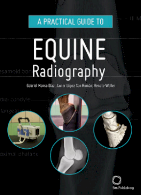
This manual is designed to serve as a guide for the clinical veterinarian to perform radiographs on horses. Every area of the horse is included, from the most common (helmet or giblet) to the most difficult (as the spine, thorax or abdomen). The text is limited to the basic steps a professional should follow to obtain each projection. While the images in each projection consist of a photograph of the patient's positioning, an X-ray example, the same X-ray highlighting the major anatomical structures and a three-dimensional image to demonstrate anatomy.
A Practical Guide to Equine Radiography is a hands-on manual on positioning and radiographic anatomy in the horse, suitable for vets and veterinary students.


